Prenatal care has come a long way past just making sure a baby is growing well. Thanks to modern imaging, fetal medicine & 3D/4D ultrasonography can allow physicians to examine every tiny detail of a baby’s growth and health in utero. These studies allow the physician to see, in detail and in real time, the baby’s body parts and movements, enabling early consideration of possible health concerns and fostering connectedness with the baby.
The 3D and 4D ultrasound technology is the one that provides the most detailed view of the fetus during pregnancy and is a vital step in prenatal care. To put it briefly, the 3D/4D-sound technique is not only a high-tech application but also a key factor in safer, more intelligent fetal care.
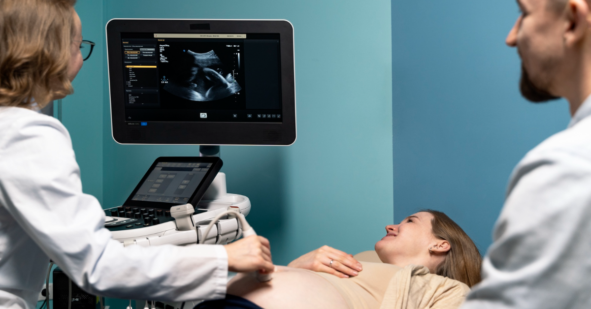
About Fetal Medicine & 3D/4D Ultrasonography
Fetal medicine is an area of obstetrics that evaluates the health and growth of the unborn fetus, which is monitored by the expectant mother and baby. A 3D/4D ultrasound is an accompanying procedure. With 3D ultrasound, moms and dads can see the baby’s lifelike images in three dimensions, while 4D captures the baby’s full-body movements. Parents and doctors can view the baby stretching, smiling, or yawning in real time!
When comparing these two advanced scans to the traditional two-dimensional ultrasound scan and flat pictures, the in-depth examination of the baby’s structures, organs, and growth patterns is greatly facilitated, which, in turn, enables early identification and management and enhances the health and well-being of both the mother and the baby.
Ways 3D/4D Ultrasonography Enhances Diagnostic Precision
Detailed Anatomical Visualization
3D/4D imaging enables the physician to examine the baby’s face, limbs, and organs, as well as more subtle aspects such as fingers and facial expressions, from various angles. This process identifies physical/structural issues (e.g., facial, cardiac, and skeletal anomalies) even earlier than conventional 2D imaging.
Real-Time Monitoring of Movement and Function
The 4D scan captures live motion. Parents have the opportunity to see their baby struggle or blink, stretch or swallow; the physician views heartbeats and baby movements whilst knowing full well that this observation of health is being added onto the routine assessment.
Better Parental Counselling and Engagement
The ability to see real, clear 3D footage of their baby provides a better platform for discourse with the parents. The physician can describe what they are seeing, clarify observations, and reduce parental anxiety. This also provides useful, dynamic notations of the fetal health that the parents can participate in.
Improved Planning for Delivery and Post-Natal Care
When doctors see potential problems with the 3D or 4D scans, they can make a delivery and special care plan if needed. The doctor may even wish to plan for which hospital to go to, alert or prepare a neonatal care team, or coordinate treatment before delivery, depending on the situation.
Enhanced Record-Keeping and Follow-Up
Digital data collected from scans can be stored and compared over time. The information this provides will allow a doctor to track growth and development patterns and assess the baby’s development during a subsequent visit. This ongoing comparison will be particularly useful during high-risk pregnancies.
Key Considerations and Optimal Use in Fetal Medicine
Timing and Indications
The optimal time for 3D/4D imaging is during the second trimester, when the features are well-formed and there is sufficient amniotic fluid for clear imaging. 3D and 4D scans are typically incorporated with regular 2D ultrasounds to provide the most complete image of the baby’s health.
Safety and Protocols
When performed properly, ultrasound is a safe, non-radiation technology. Medical practitioners have advised that a 3D or 4D scan should be used strictly for medical purposes, rather than just for a nice video as a keepsake or for enjoyment. Herein, in the standard ultrasound imaging scenario, the physician’s skill and the machine’s quality also affect the results’ precision.
Integration with Other Diagnostic Tools
3D and 4D scans provide clear images, but they do not replace other parts of an image evaluation. Practitioners frequently integrate them with other clinically focused tools, such as Doppler studies, MRI, or genetic testing, to provide a more complete understanding of the baby.
Cost and Accessibility
To get hold of the specialized machines and trained professionals for 3D and/or 4D ultrasound, one has to pay a price much higher than that of traditional ultrasound. A lot of fetal medicine clinics would suggest the use of 3D or 4D ultrasound in a high-risk pregnancy situation or when the doctor needs clear views to take medical decisions or plan interventions.
Emotional and Technical Support
Witnessing the first glimpse of your baby’s face can generate a lot of emotions. Sometimes the results can be mixed. A caring fetal medicine team provides guidance to parents regarding the scan findings, answers their questions, and provides appropriate care throughout.
Conclusion
The combination of Fetal Medicine & 3D/4D Ultrasonography has transitioned the way physicians practice regarding caring for unborn babies. We are now able to diagnose problems earlier, plan treatment sooner, and help parents connect with their baby in a unique and special way.
If you’re looking for advanced fetal imaging and comprehensive prenatal fetal medicine support, Sunshine Women’s Hospital offers expert-led 3D/4D ultrasonography in a compassionate, high-tech setting.
Our fetal medicine specialists will ensure that every mother leaves with clear explanations and personalised care from the earliest stages of pregnancy.
Contact Sunshine Women’s Hospital today to schedule your 3D/4D ultrasonography, and take your first step to a safer and more confident pregnancy journey.